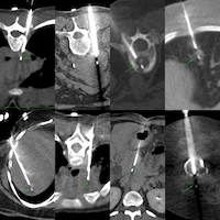
Case 162: Subgaleal Calvarial Lesion Biopsy
New Onetime Lifetime Subscription

Current Case:
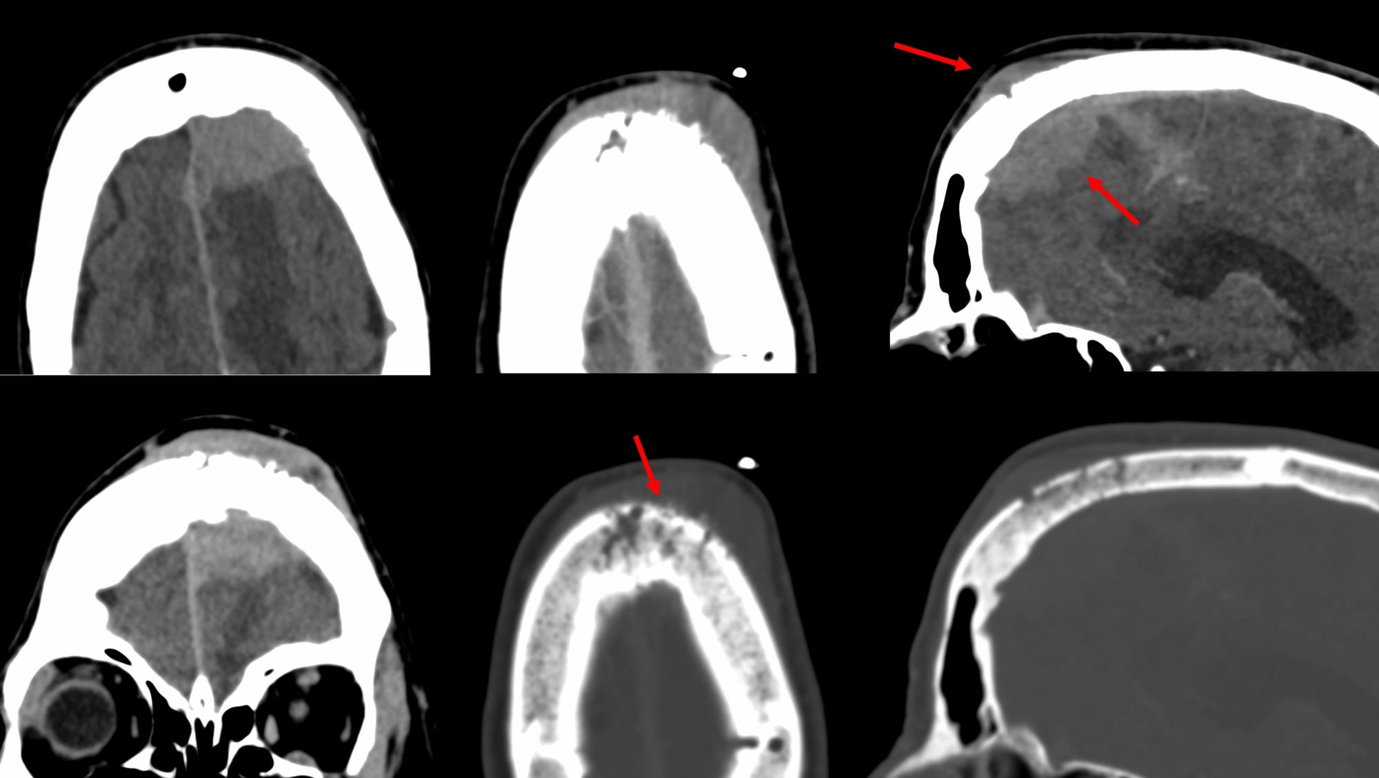
A 70-years old presented with left frontal swelling and headache.
She was posted for a biopsy of the subgaleal soft tissue, but she did not turn up. 2 months later, she presented with progression of disease.
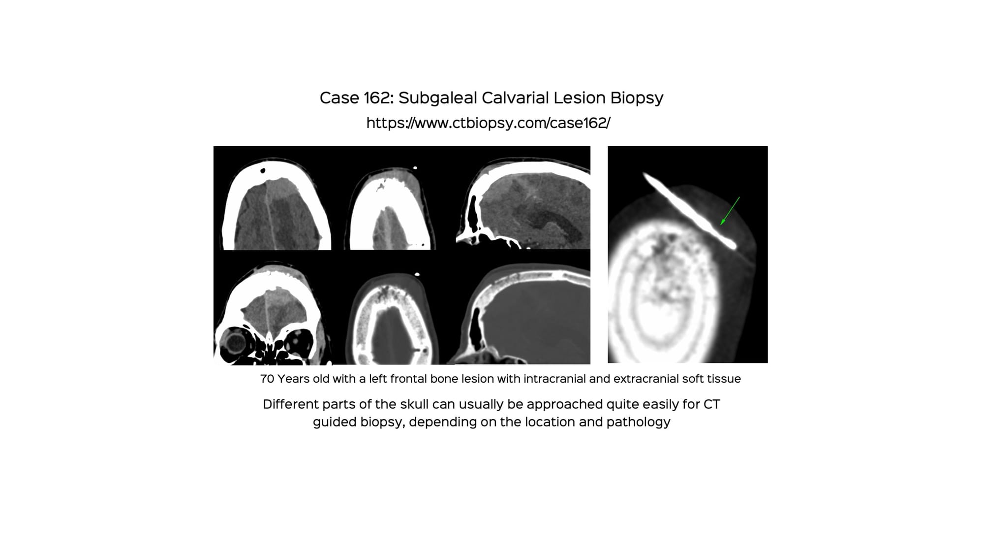
The video describes the case and the approach with 2 other examples of different skull and calvarial biopsies.

Sign up for CT Guided Biopsy
Understanding, learning and discussing CT guided biopsies and ablations.
No spam. Unsubscribe anytime.
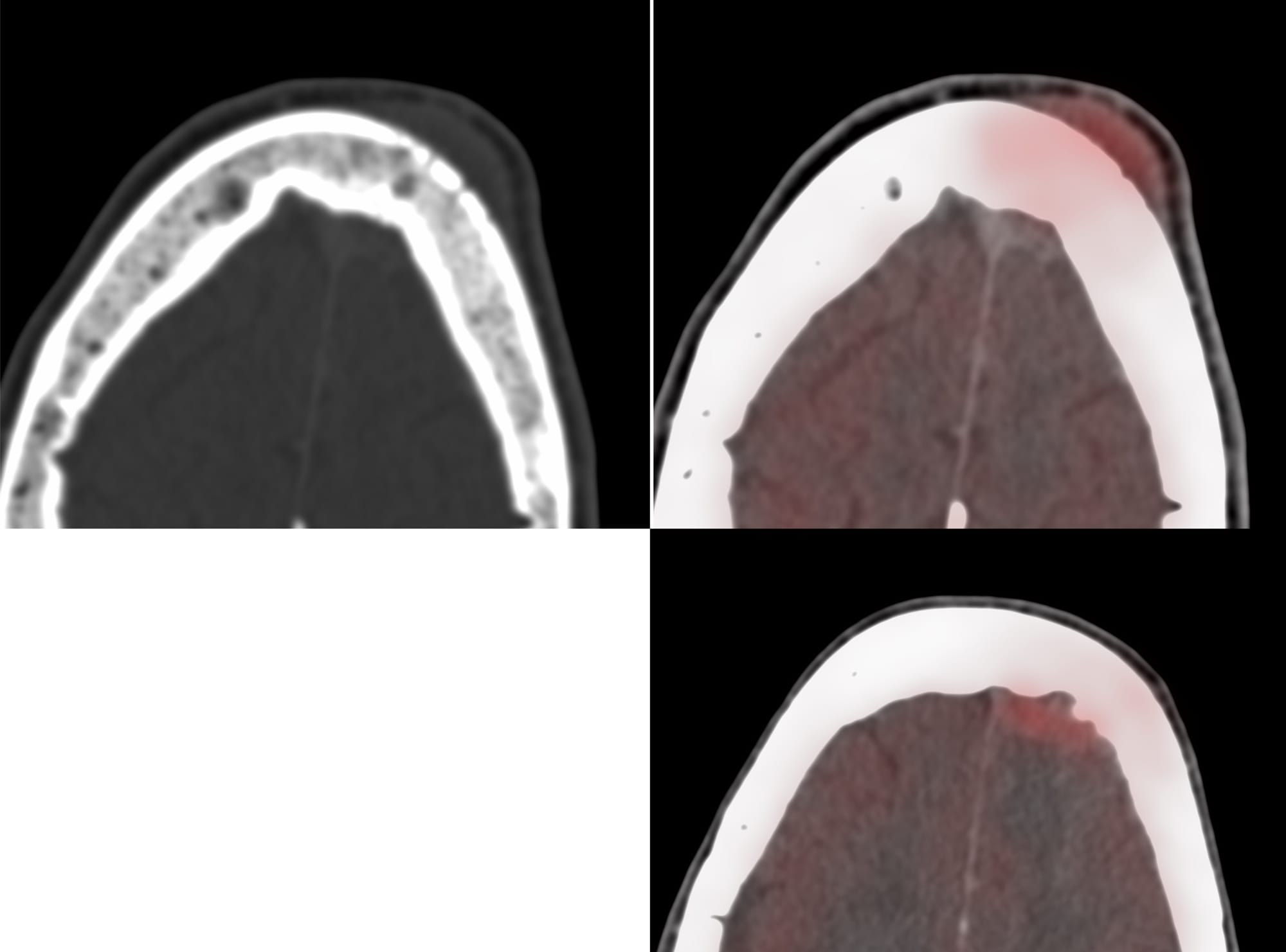
Region: Skull, Calvarium
Age: 70
Findings: Intra and extradural lesion with permeative frontal bone involvement
Lesion Biopsied: Subgaleal calvarial soft tissue
Size of Lesion: Not relevant
Gun: 18G Cook, 10 mm throw, short
No of cores: 11 for histopath in 5 vials and four for microbiology
Sedation: Conscious
Position & Approach: Oblique, out-of-plane
Time Taken (marker to wash-out): 11 mins
Complication: None
Level of Difficulty: 3/5
Diagnosis: Non-Hodgkin’s lymphoma of follicle cell phenotype