bjankharia | Instagram, Facebook | Linktree
Radiologist, Writer, Atmasvasth

Current Case:

A 54-years old with a mesenteric mass presented for a biopsy - the mass was unchanged over 6 months.

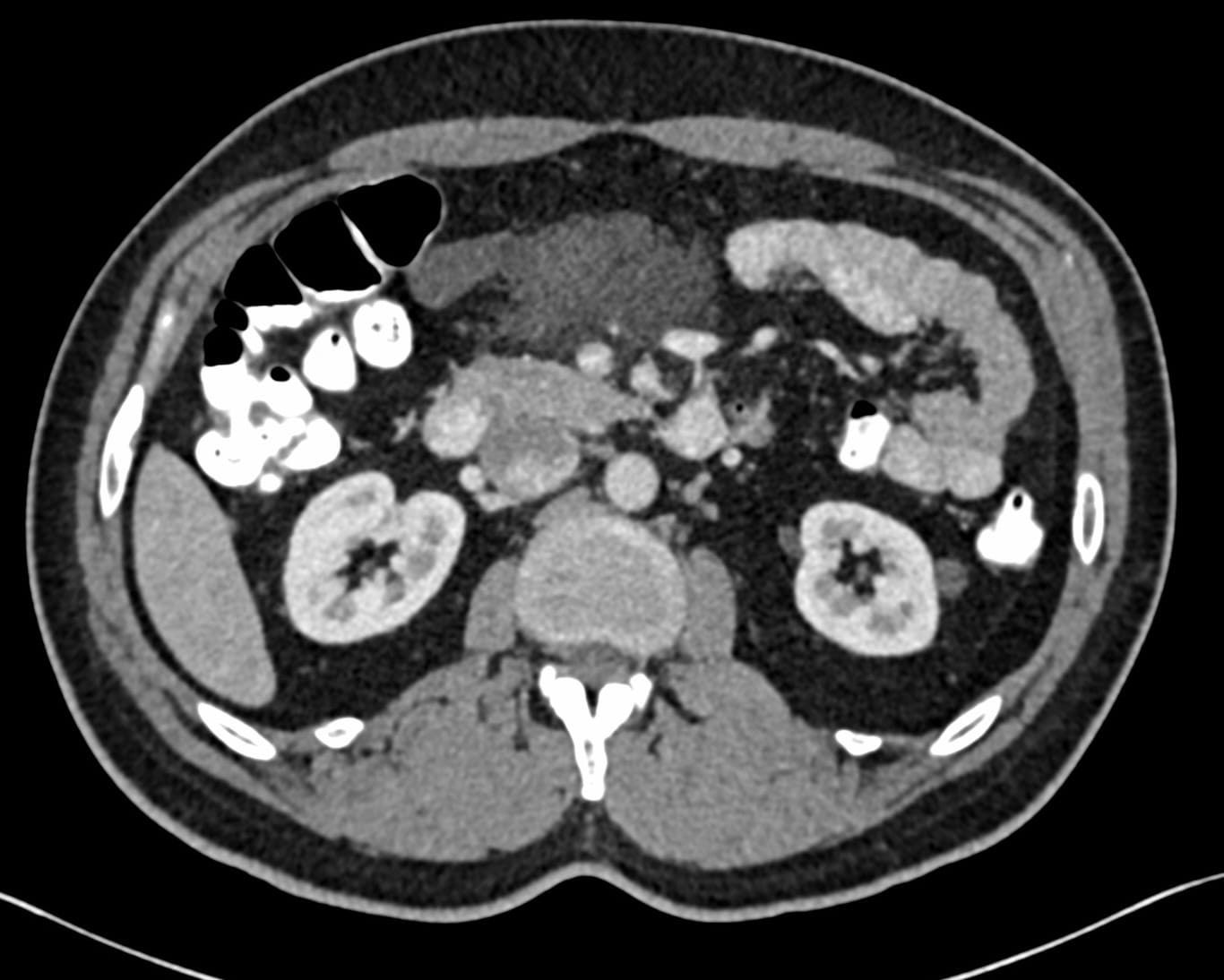
Though, this was likely a lymphatic cyst, the surgeon wanted a biopsy, because one of the earlier reports had raised the possibility of lymphoma.

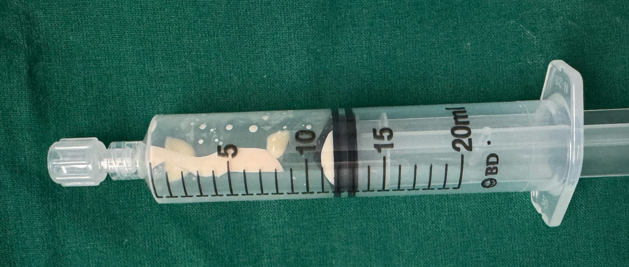
The biopsy was performed with an 18G BARD coaxial gun. Milky fluid was aspirated, which confirmed the diagnosis.

The video discusses the case, the approach to this lesion and a discussion of cases where a diagnosis can be made on table due to the look of the material or the feel during biopsy.

Please note that the video only plays within this browser page and not outside of this browser page. If the video does not autoplay in your email, then you will need to open this page in your browser, by clicking "View Online", in the 2nd line below the title of the post and next to my name.

Region: Mesentery
Age: 54 yrs
Findings: Mesenteric cystic lesion
Lesion Biopsied: Mesenteric cystic lesion
Size of Lesion: -
Gun: 18G BARD, 20 mm throw, long
No of cores: 4 for histopath and milky aspirate for microbiology and triglyceride levels
Sedation: No
Position & Approach: Supine
Time Taken (marker to wash-out): 9 mins
Complication: None
Level of Difficulty: 2/5
Diagnosis: Mesenteric lymphatic cyst

Table of Contents:

Table of Contents
Table of Contents

Previous Post:

Lecture: The Role of Image-Guided Biopsy - When the Biopsy Makes the “World of a Difference”
All bone and spine lesions have to be biopsied, unless the lesion cannot be reached safely or the patient needs emergency surgery

Other Sites and Cases:

Copy 46: A Scapular Enigma
61-yrs old treated for carcinoma with scapular lesion
Snippet 19: The Various Uses of Prone Imaging
The many uses of prone imaging during CT chest examinations