bjankharia | Instagram, Facebook | Linktree
Radiologist, Writer, Atmasvasth

Current Case:

A 67-years old presented with a fibrosing ILD in 2018, which was diagnosed as probable UIP/IPF

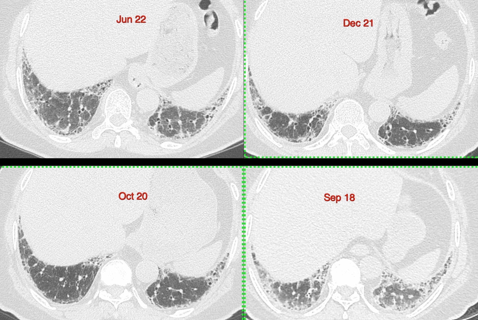
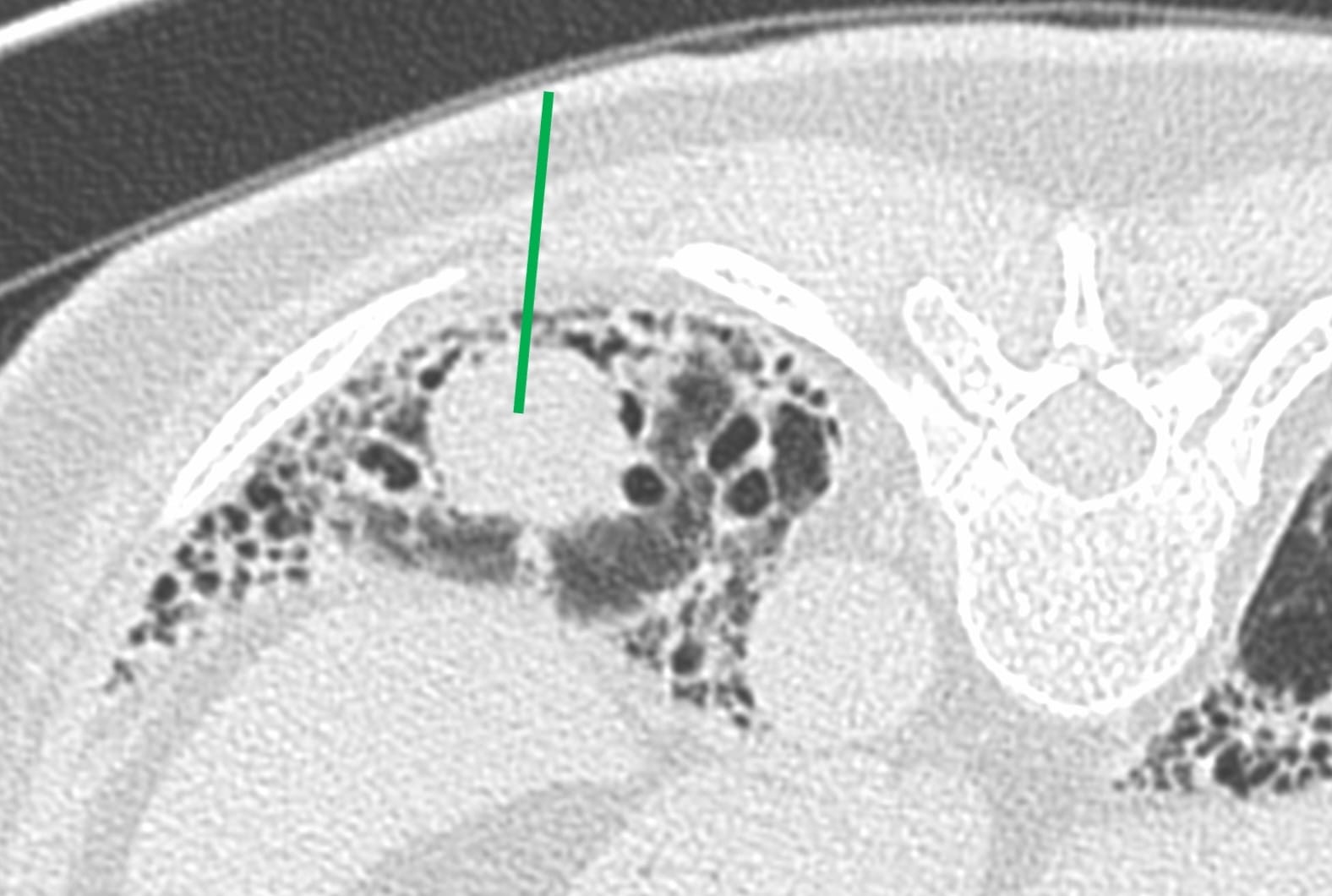
In May 24, a follow-up CT showed progression of disease and a new lung nodule.

The pulmonologist wanted a biopsy. I thought this would be the best route.

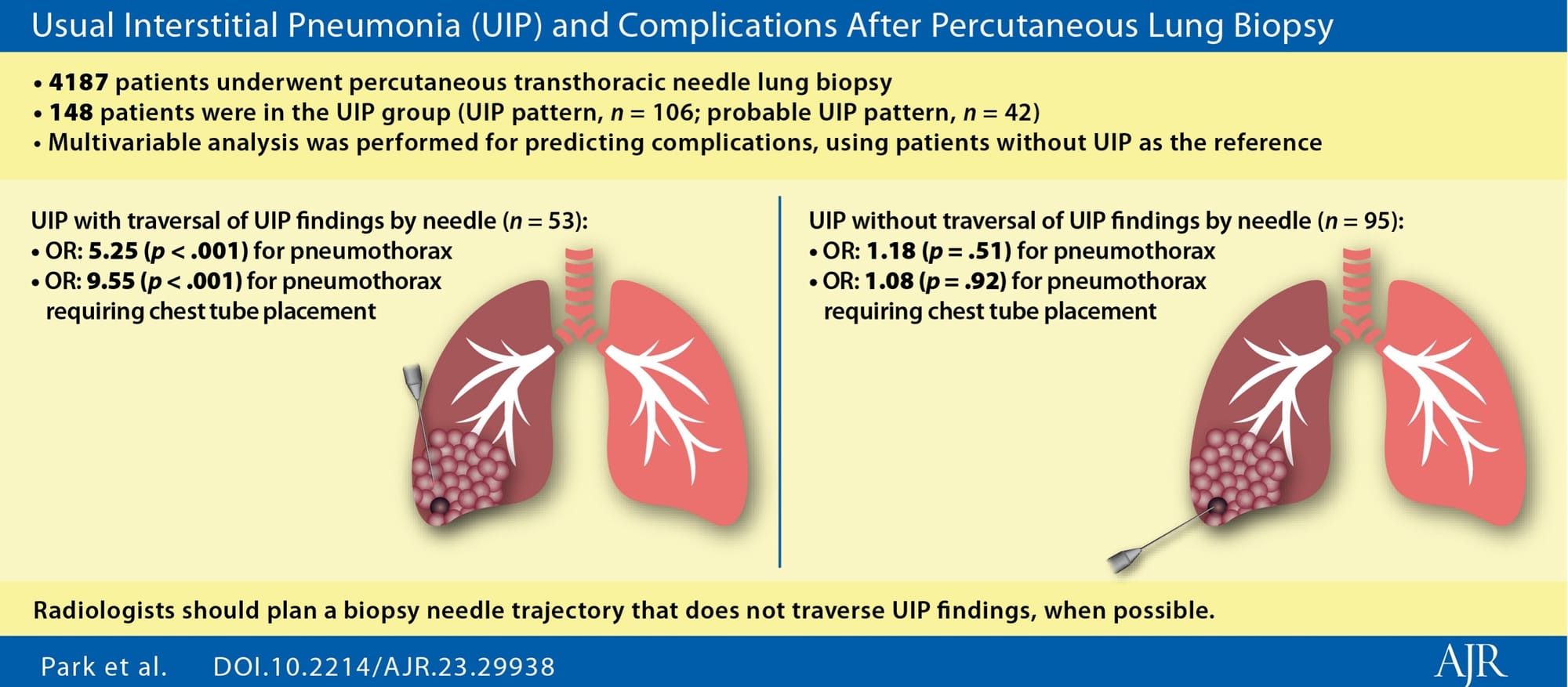
A new paper also discusses this issue.

The video discusses the case, the approach to this lesion with a discussion on biopsy of focal lung lesions in patients with fibrosing ILDs with multiple other examples

Please note that the video only plays within this browser page and not outside of this browser page. If the video does not autoplay in your email, then you will need to open this page in your browser, by clicking "View Online", in the 2nd line below the title of the post and next to my name.

Region: Lung
Age: 73
Findings: New lung nodule in a UIP/IPF background
Lesion Biopsied: Nodule
Size of Lesion: 20.8 mm z axis x 20.22 mm - avg size - 20.5 mm
Gun: 20G Cook, 10 mm throw, short
No of cores: 6 for histopath and 2 for microbiology
Sedation: No
Position & Approach: Prone
Time Taken (marker to wash-out): 8 mins
Complication: None
Level of Difficulty: 4/5
Diagnosis: Granulomatous disease, negative GeneXpert

Table of Contents:

Table of Contents
Table of Contents

Similar Post:

Case 15: UIP IPF with a Superimposed Lung Nodule
75-years old man with UIP IPF and an associated lung nodule that was biopsied - squamous cell carcinoma.

Previous Post:

Lecture: The Role of Image-Guided Biopsy - When the Biopsy Makes the “World of a Difference”
All bone and spine lesions have to be biopsied, unless the lesion cannot be reached safely or the patient needs emergency surgery

Other Sites and Cases:

Case 100: CT of Covid-19...4 Years and 3 Months Later
The many faces of Covid-19, 4 years later