bjankharia | Instagram, Facebook | Linktree
Radiologist, Writer, Atmasvasth

Current Case:

A 66-years old with both metastatic neuroendocrine tumor (NET) and metastatic papillary renal carcinoma came with a lesion on the caudate lobe surface possibly involving it.

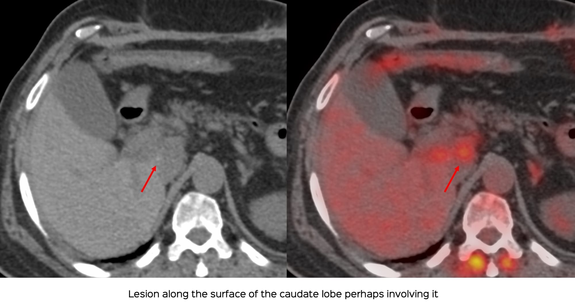
The oncologist wanted a biopsy to differentiate between renal cell and NET metastasis.

Which route will you take?

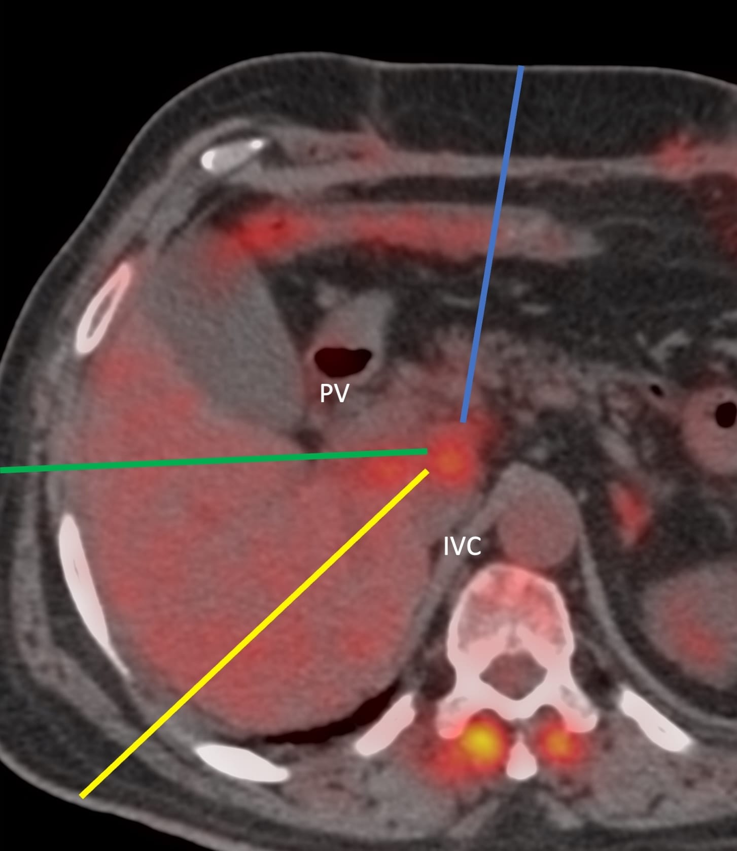
The video discusses the case, the approach to this lesion and a discussion on different transorgan approaches.

Please note that the video only plays within this browser page and not outside of this browser page. If the video does not autoplay in your email, then you will need to open this page in your browser, by clicking "View Online", in the 2nd line below the title of the post and next to my name.

Region: Liver
Age: 66 years
Findings: Lesion on surface of an involving caudate lobe
Lesion Biopsied: Surface caudate lobe liver
Size of Lesion: 27.4 mm z axis x 18.7 mm
Gun: 20 G Cook
No of cores: 8 for histopath in 6 vials
Sedation: No
Position & Approach: Supine, transpancreatic
Time Taken (marker to wash-out): 14 mins
Complication: None
Level of Difficulty: 4/5
Diagnosis: Metastatic renal cell carcinoma

Table of Contents:

Table of Contents
Table of Contents

Previous Post:

Lecture: The Role of Image-Guided Biopsy - When the Biopsy Makes the “World of a Difference”
All bone and spine lesions have to be biopsied, unless the lesion cannot be reached safely or the patient needs emergency surgery

Other Sites and Cases:

Case 99: A Cleft Lung Puzzle
50-yrs old with an unusual but otherwise straightforward diagnosis
Case 45: Shoulder in Ruin
53-yrs old with pain and swelling in the left shoulder