
This 78-years old man came for a nasopharyngeal mass biopsy to get material for microbiology. The head and neck surgeon was not comfortable doing this biopsy and so the infectious disease physician asked for a CT guided biopsy.
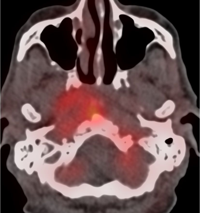
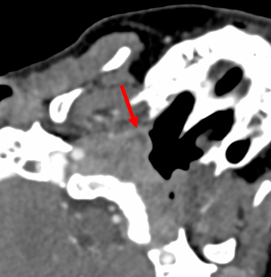
At the time of the biopsy, the scan showed necrosis / collection anteriorly.
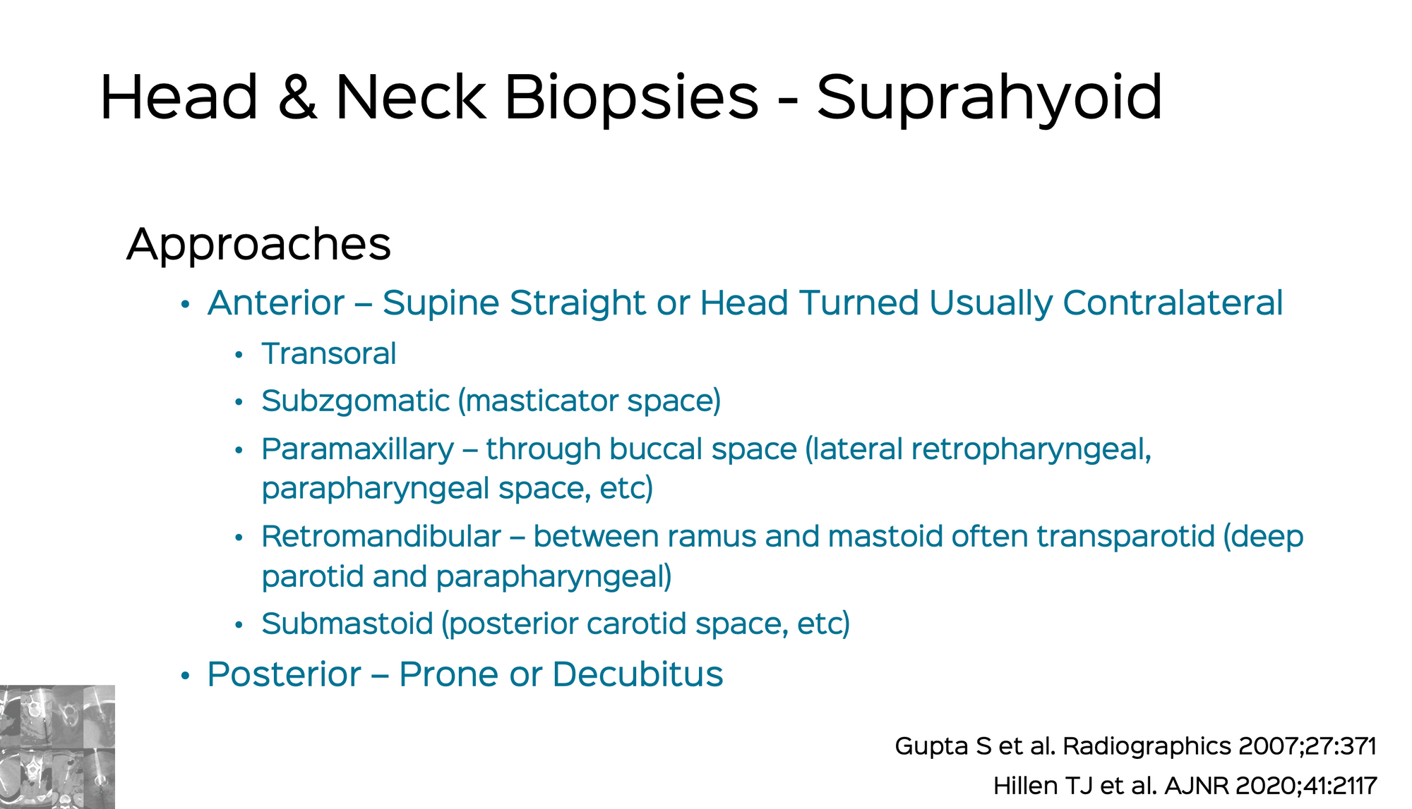
Which route will you take?
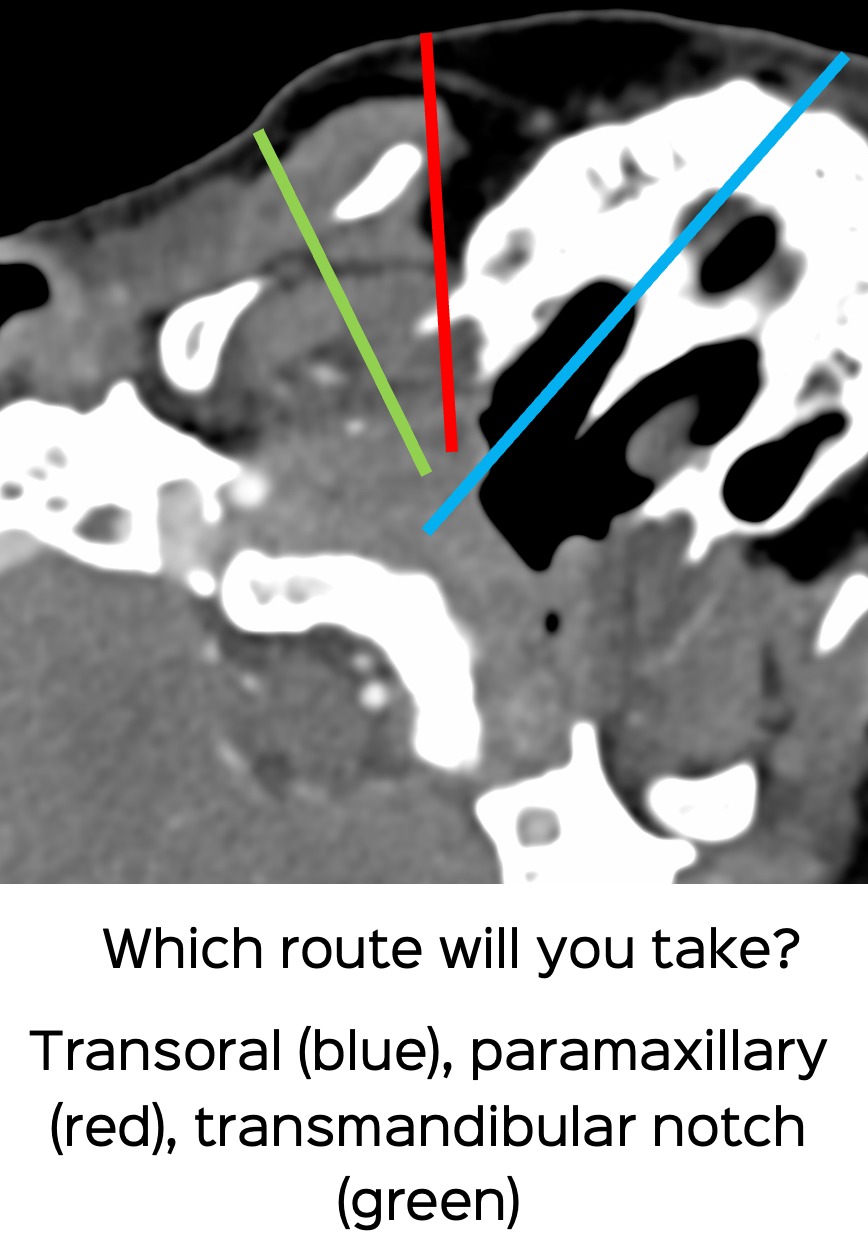
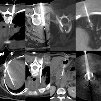
These are the typical routes available for reference
These are some of the previous head and neck biopsies discussed on the site for reference.
Case 23: Biopsy of Greater Wing of Sphenoid in a 25-Years Old for Recurrent Adenoid Cystic Carcinoma
25-years old treated with h/o adenoid cystic carcinoma palate with intra-orbital mass involving sphenoid bone, which was biopsied.
 Bhavin Jankharia
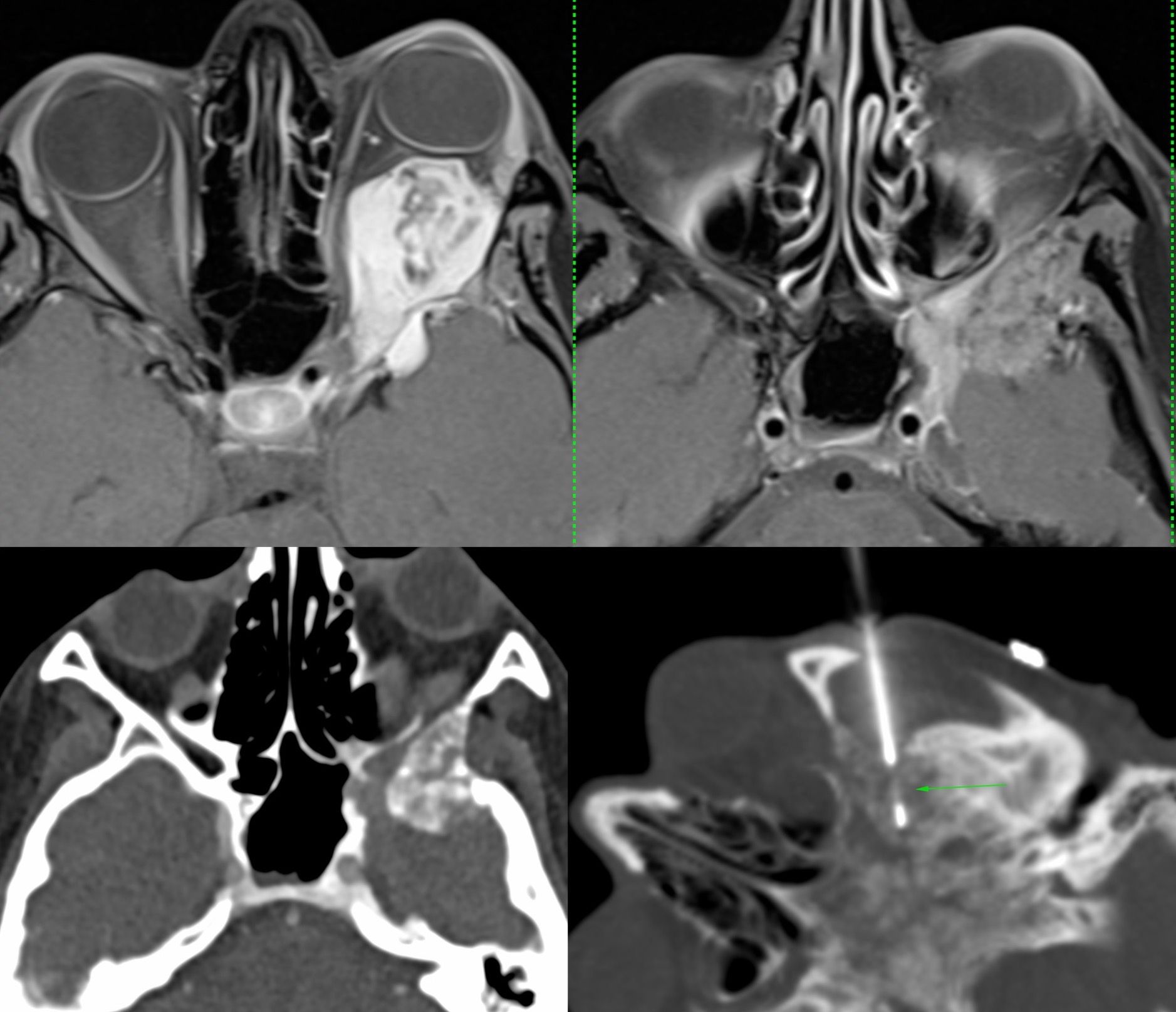
Case 24: Oropharyngeal / Parapharyngeal Mass Biopsy
68-years old with a left parapharyngeal- oropharyngeal mass. The CT guided biopsy was simple. The video explains safe routes and discusses two papers.
Bhavin Jankharia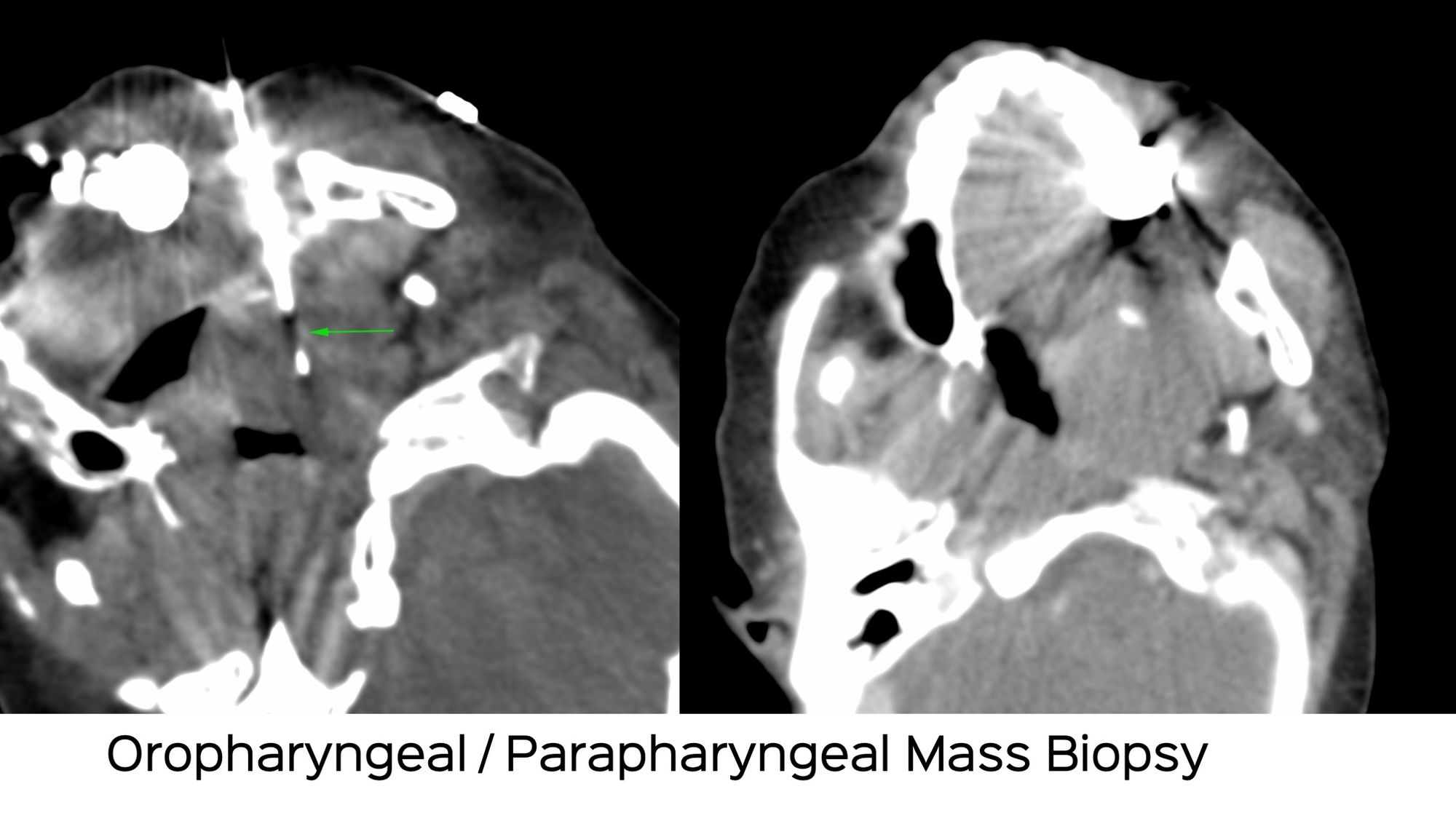
Case 78: Lateral Pharyngeal / Retropharyngeal Lesion Biopsy with a Paramaxillary Approach
Lateral Pharyngeal / Retropharyngeal Lesion Biopsy with a Paramaxillary Approach
Bhavin Jankharia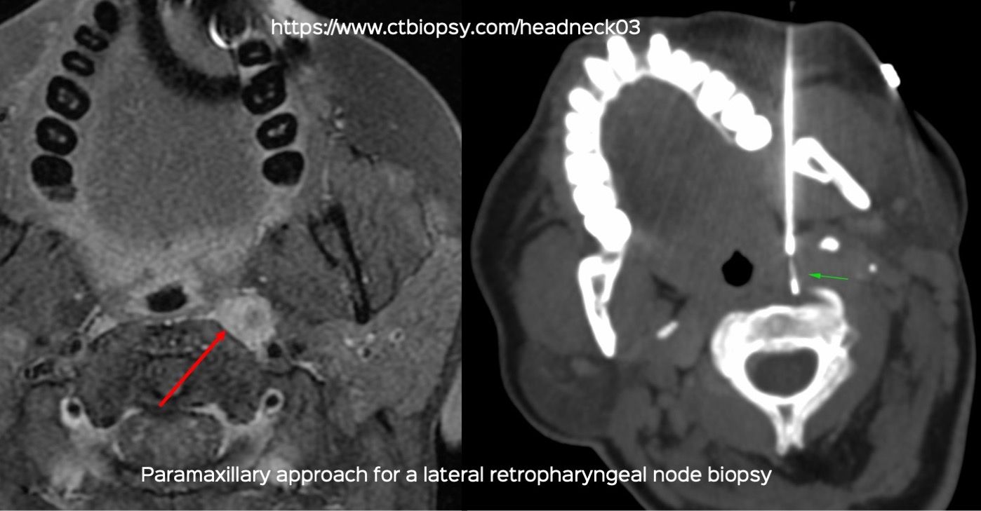
What would you do?
The discussion video follows.